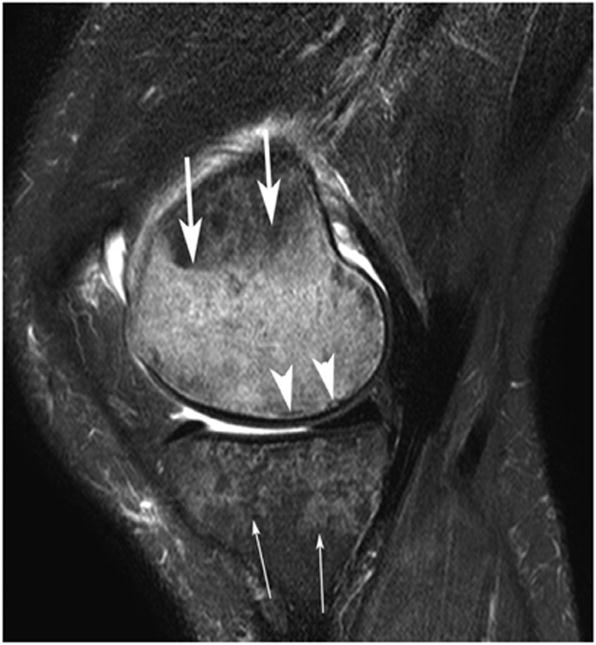
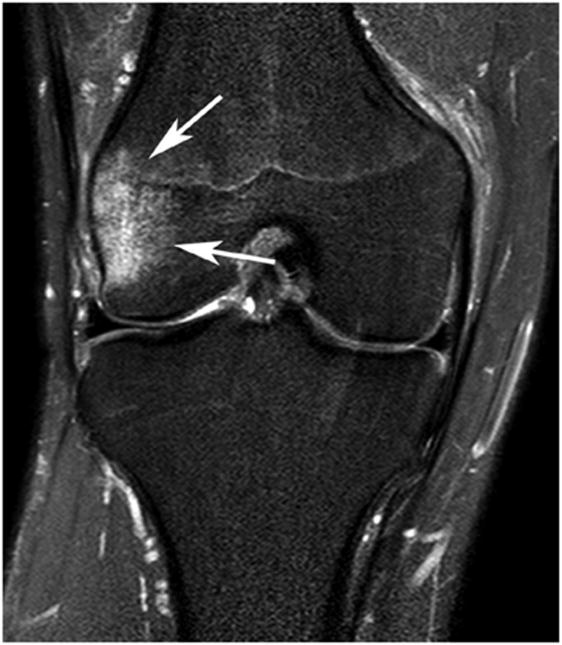
Causes of subchondral signal alterations of the knee joint, not due to osteoarthritis.
(a) Subchondral Insufficiency Fracture - Sagittal IW fat-suppressed MRI shows a subtle subchondral irregular hypointense line (arrowheads) of the medial femoral condyle which represents subchondral insufficiency fracture surrounded by an extensive hyperintensity bone marrow edema (arrows). Also there is a large heterogeneous hyperintensity of the medial tibial plateau (thin arrows) typical for osteoporosis in this 59-year-old woman. Note there is a small joint effusion.
(b) Traumatic Bone Marrow Secondary to Transient Lateral Subluxation of the Patella - Sagittal IW fat-suppressed MRI shows moderate hyperintensity of the lateral femoral condyle (arrows) distant from the subchondal bone and typical for traumatic bone marrow secondary to transient lateral subluxation of the patella. Note there is also a grade 1 medial collateral ligament sprain


