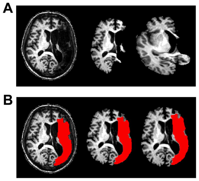
Figure 1.

Example of using the lesion mask for the brain extraction and normalization steps. (A) Standard brain extraction of lesioned anatomical MRI (left) results in removal of viable tissue beyond the stroke lesion (middle) and subsequent misregistration to standardized space (right). (B) The lesion mask (in red) was traced on each subject’s anatomical MRI (left) and applied to achieve good results during the brain extraction algorithm (middle) and subsequent normalization to standardized space (right). Images are presented in radiological convention (left=right).
