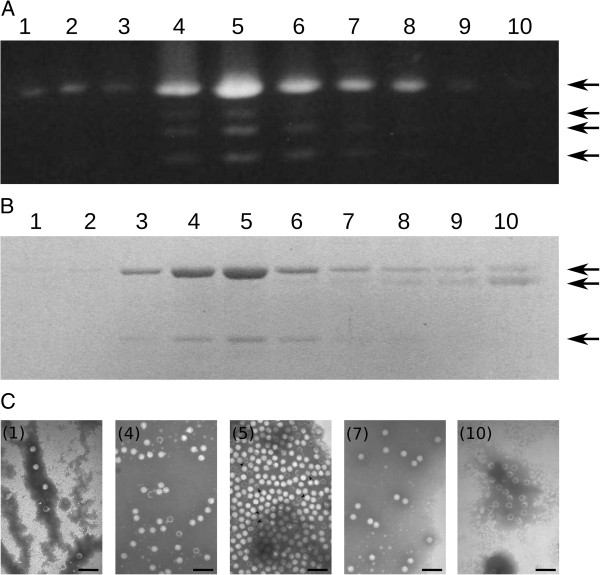
Figure 2.
Analysis of the gradient fractions from particulate material of B. cinerea CCg378, using 0.8% (w/v) agarose gel electrophoresis and 10% (w/v) SDS-PAGE. (A) Nucleic acids present in gradient fractions 1–10. (B) Polypeptide profile of gradient fractions 1–10. The arrows on the right indicate the migration position of dsRNAs (A) and major polypeptides (B). (C) Electron micrograph of mycoviruses from B. cinerea CCg378 present in representative gradient fractions. Viral particles of gradient fractions 1, 4, 5, 7 and 10 are shown. Arrows in C (5) indicate the position of some mycoviruses of 23 nm in diameter. Negative staining with 2% (w/v) potassium phosphotungstate, pH 7.0. The bar marker in 1, 4, 5, 7 and 10 represents 100 nm.

