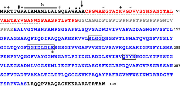
Figure 1.

Primary structure of the CvChi45 protein encoded by the CV2935 ORF of C. violaceum ATCC 12472. The amino acid sequence of CvChi45 is shown, highlighting the N-terminal signal peptide (underlined with a continuous line), the ChBD (in red), the Pro/Thr-rich linker (in gray), the CatD (in blue) and the C-terminal extension (in black). The central h-region of the signal peptide (SP) is indicated by a gray bar. The symbols above the SP sequence refer to positively (+)-charged residues in the n-region (before the h-region), Gly residues (#) flanking the h-region, and Ala residues (●) in the c-region (after the h-region). The boundaries between these regions were determined by the SignalP 3.0 program [66]. The N-terminal sequence that was experimentally determined for the recombinant protein, which was purified from the culture medium of induced E. coli cells, is underlined with a dashed line. The actual SPase I cleavage site is indicated by an arrow. Positively (+)- and negatively (-)-charged residues within the first twenty N-terminal residues of the mature protein are also indicated. Structural motifs in the CatD that are involved in substrate binding and catalysis are boxed, and the crucial catalytic Glu residue is indicated by an asterisk (*). The numbers of the residues relative to Met1 are shown on the right side of the sequence.
