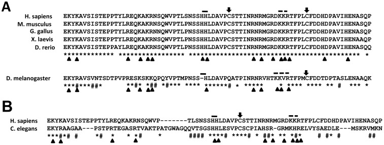
Figure 2. Sequence conservation of the putative DNA binding domain (DBD) of vertebrate INI1/hSNF5, Drosophila SNR1 and C. elegans SNF5.
(A) Sequence comparison of the putative DNA binding domain of Drosophila SNR1 with the domains in vertebrates. Conserved residues are shown by (*) and partially conserved residues by (#). The HH and KKR motifs are shown with dash and broken dash, respectively. The conserved cysteine residues are shown by an arrow. The conserved lysine, arginine and histidine residues are shown with an arrowhead. (B) Sequence comparison of the putative DNA binding domain of C. elegans SNF5 with the human domain.

