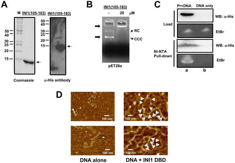
Figure 3. DNA binding studies of INI1 DBD.
(A) Coomassie blue stained SDS-PAGE (left panel) and Western blot analysis (right panel) of recombinant, purified INI1 DBD using α-His antibodies as probes. (B) Agarose gel retardation assay (AGRA) of INI1 DBD using 100 ng of pET28a as substrate and indicated amount of polypeptide; RC: relaxed circular DNA, CCC: covalently closed circular DNA. (C) Ni-NTA pull-down assay of INI1 DBD:U5 HIV-1 LTR complex (lane a) and of DNA alone (lane b). The precipitated complex was analyzed by western blot using α-His antibodies as probes to detect INI1 DBD and the co-precipitated DNA was analyzed by ethidium bromide (EtBr) staining following 10% urea-PAGE. The loading controls are shown. (D) Atomic force microscopy (AFM) images of pNEB206A DNA alone (left panel) and in complex with INI1 DBD (right panel). Regions of DNA coated with protein (arrows) and free DNA (arrowhead) is shown.

