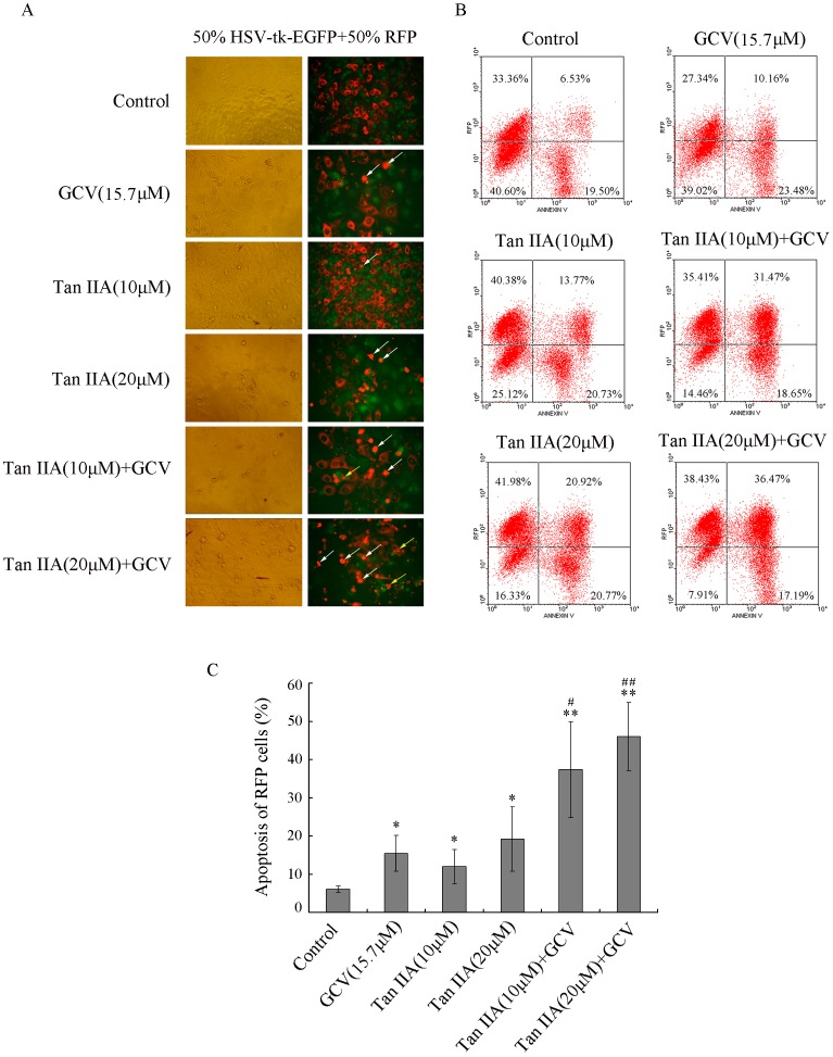
Figure 3. Tan IIA enhances the bystander effect.
Stable HSV-tk-EGFP and RFP B16 cell lines were mixed at a ratio of 1∶1. The mixture of cells was left untreated or treated with Tan IIA (10 or 20 µM) for 24 h, and then was cultured with or without GCV (15.7 µM) for 48 h. (A) Representative images as shown by fluorescence microscopy. The red fluorescence in living RFP cells mainly localizes in cytoplasm; the aggregation of red fluorescence indicates cell shrinkage which is the hallmark of apoptosis (white arrows); the clustered light spots indicate the formation of apoptosis bodies (yellow arrows). (B) The apoptosis of RFP cells was analyzed by flow cytometry with annexin V stain. (C) Quantification of three independent experiments. *p<0.05, **p<0.01 compared with control; #p<0.05, #p<0.01 compared with Tan IIA or GCV treatment alone.

