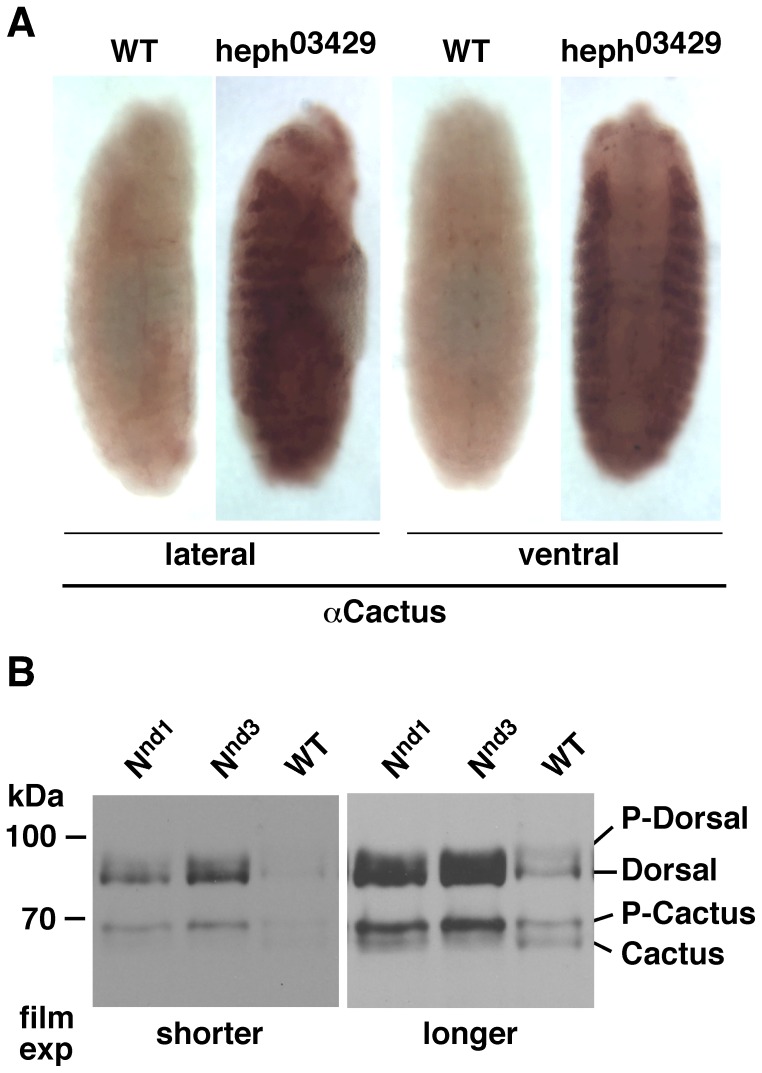
Figure 1. P-Cactus and P-Dorsal levels in embryos that over-express Notch globally.
A. Cactus accumulates to a high level in the lateral regions of heph03429 embryos. Wild type (wt) strain used here and throughout the study is the yellow white (yw) strain. Embryos shown are at stages 13–14 probed with the Cactus antibody. All staging in this study was done according to [49]. B. Nnd1 and Nnd3 embryos express higher levels of P-Cactus in association with higher levels of Dorsal (the unphosphorylated, cytoplasmic form) [43]–[46]. Two different exposures to film are shown for clarity. The same blot was probed with the two different antibodies. Eggs of all genotypes were collected at room temperature at 1–3 hour intervals and incubated at 30°C for 1–3 hours before protein extraction. The same result was obtained from all samples. Data for 3-hour collection and 1-hour 30°C incubation are shown. The same number of embryos was used in each extraction and the same amount of the sample was loaded in each lane. Immuno-labeling experiments were repeated twice and most embryos of each stage showed the same phenotype. Western blotting experiments were repeated three times.

