
Figure 3. No EpoR detected by A82 or M-20 in normal and cancerous human breast tissues (Her2+ and Her2−) but M-20 detected a 59 kDa band in MCF-7 Cells.
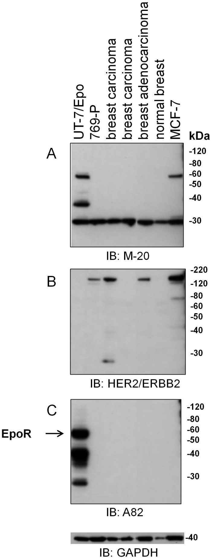
EpoR protein expression analysis was performed by immunoblotting (IB) with A82 and M-20. Four identical immunoblots with the same master mix were performed at the same time and probed with different antibodies under the same conditions. Lysates included tissue samples from 3 different human tumor biopsies, a normal breast sample or MCF-7 cells. The positive control was UT-7/Epo cells and the negative control was 769-P cells. The positions of molecular weight markers are shown. (A) Anti-EpoR monoclonal antibody M-20 (lot E2004). (B) Anti-Her2 antibodies. (C) Anti-EpoR antibody A82. A blot with anti-GAPDH antibodies served as a loading control.