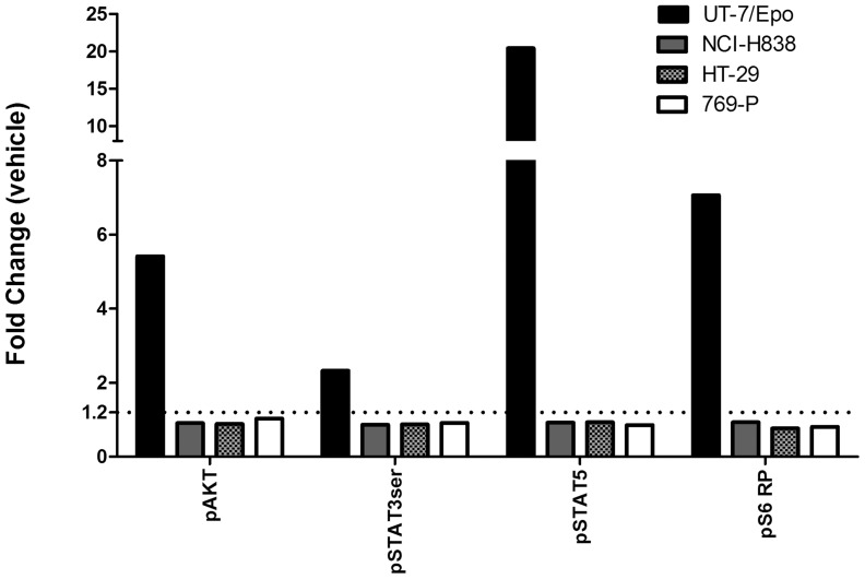
Figure 9. No effect of Epo on pAKT, pSTAT3, pS6RP or pSTAT5 with NCI-H838 lung carcinoma cell line by Phosflow.
Cells were grown, treated for 30 minutes with the indicated growth factors and recovered. Fixed and permeabilized cells were washed and stained at room temperature with fluorochrome-conjugated antibodies specific for the phosphorylated (p) forms of STAT5, STAT3 and AKT, and S6 ribosomal (S6RP) proteins. Stained cells were run on a LSRII (FACS) instrument. For both vehicle and Epo treated samples 10,000 events were acquired during the FACS acquisition. Through gating and use of caspase 3 all events were from viable and intact cells. The data in the figure represents a single measurement for each cell line across all measured phospho-proteins. Results are reported as mean fluorescence intensity (fold change) in treated samples compared to vehicle. The dotted line shows the threshold level of staining that can be detected above background. This value represents the minimum fold change relative to vehicle required to accurately define a positive cytokine response signal in the Phosflow assay. The threshold fold change of 1.2 was experimentally validated previously through experiments where stimulated cells were titrated into negative control cells (data not shown). UT-7/Epo cells were the positive control and 769-P and HT-29 cells were negative controls.

