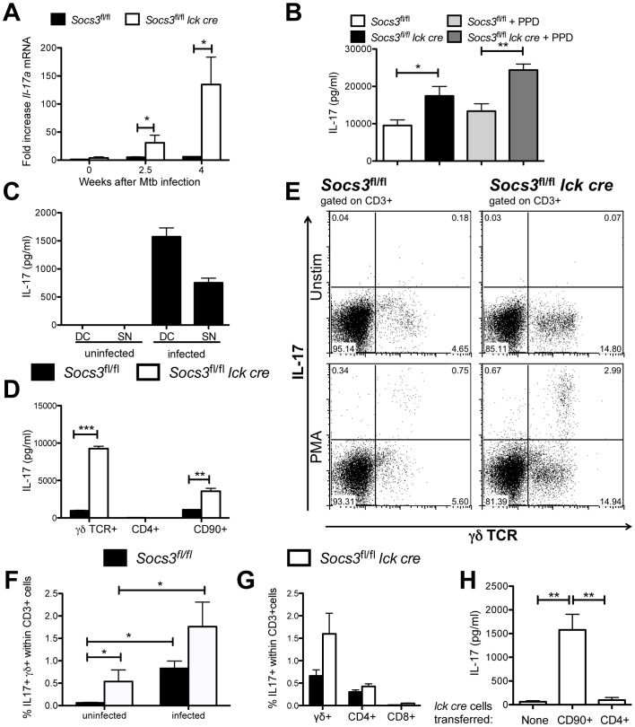
Figure 7. SOCS3-deficient γδ+ T cells secrete IL-17 during M. tuberculosis infection.
Socs3fl/fl lck cre and Socs3fl/fl mice were sacrificed before and at 2.5 and 4 weeks after M. tuberculosis infection and the total RNA was extracted from lungs. The accumulation of Il-17a and Hprt transcripts was measured by real time PCR (A). The mean fold increase of IL-17a mRNA ± SEM in lungs from infected mice (n≥5 per group) was calculated. One out of two independent experiments is depicted. Differences with infected Socs3fl/fl mice are significant (*p<0.05 Student t test). Socs3fl/fl lck cre and Socs3fl/fl mice were sacrificed 2.5 weeks after aerosol infection with M. tuberculosis. Lung cell suspensions were stimulated or not with 20 µg/ml PPD for 48 h. The IL-17 level in supernatants was determined by a cytokine bead assay (CBA) (B). The mean IL-17 concentration ± SEM (n≥6 animals per group) is depicted. Differences in cytokine concentrations are significant (*p<0.05, **p<0.01 ANOVA with Bonferroni correction). CD90+ naïve spleen T cells were co-cultured either with uninfected, BCG-infected BMDCs (DC) or with their 48 h supernatants (SN). After 72 h, the IL-17 levels in culture supernatants were measured by ELISA. A representative out of three independent experiments is shown (C). 105 γδ+, CD4+ or CD90+ FACS sorted T cells from Socs3fl/fl lck cre and Socs3fl/fl mice were co-cultured with supernatants from BCG-infected BMDCs for 72 h. The mean IL-17 concentration in supernatants from triplicate cultures ± SEM is depicted (D). Differences in cytokine concentrations are significant (**p<0.01, ***p<0.001 Student t test). The presence of IL-17-secreting cells in PMA/ionomycin-stimulated lung cell suspensions from Socs3fl/fl lck cre or Socs3fl/fl mice before or 16 days after infection with M. tuberculosis was measured by FACS as described in materials and methods. Representative FACS dot plots from CD3+ gated infected lung cells before or after PMA/ionomycin stimulation are shown (E). The frequency of IL-17-secreting γδ+ within CD3+ cells in uninfected or infected mice is displayed (n = 6, *p<0.05 Mann Whitney U test) (F). The mean frequency of IL-17-secreting CD4+, CD8+ and γδ+ within CD3+ cells in lungs of infected mice (5 mice per group) ± SEM is depicted (G). Rag1 −/− mice were infected with M. tuberculosis 2 weeks after inoculation with either 1.2×10⁶ CD4+ or 2×10⁶ CD90+ Socs3fl/fl lck cre spleen cells. Mice were sacrificed 4 weeks after infection and lung cell suspensions incubated for 48 h. The mean concentration of IL-17 in supernatants ± SEM (n = 6) is depicted (H). Differences in cytokine concentrations are significant (**p<0.01 Student t test).

