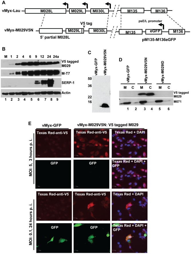
Figure 5. M029 is expressed as an early/late viral protein, associated with virions and is localized in the nucleus and cytoplasm of virus-infected cells.
A) Construction of vMyx-M029V5N. A plasmid having N-terminal V5 sequence –tagged M029 was constructed by PCR. The final recombination plasmid has 5′ partial M028L sequence, V5-tagged M029 sequence and M030L sequence and was constructed by three-element recombination of the Multisite Gateway system. The virus also has a eGFP expression cassette inserted at the intergenic region between M135-M136 locus; B) M029 protein is expressed as an early/late gene product in infected rabbit cells. RK13 cells were left uninfected (M) (lanes 1) or were infected with vMyx-M029V5N at an MOI of 3. Cells were collected at1, 2, 4, 6, 9, 12, and 24 (lanes 2 to 8) and 24a in the presence of AraC (lane 9) h p.i. The membranes were first probed with anti-Serp-1 antibody, stripped and probed for M-T7 and actin (loading control). C) M029 is packaged into MYXV virions. Gradient purified vMyx-GFP (lane 1) and vMyx-M029V5N (lane 2) viruses were separated on 12% SDS-PAGE gels for Western blotting. V5-tagged M029 was detected using an anti-V5 antibody. D) M029 is located in the membrane and core of the MYXV virion. Gradient purified vMyxGFP (lanes 1 and 2), vMyx-M029V5N (lanes 3 and 4) and vMyx-M029ID (lanes 5 and 6) were treated with detergent and DTT to separate the membrane (M) and core components (C) of the virions. The fractions were separated on SDS-PAGE for Western blotting, followed by detection using an anti-V5 antibody, stripped and probed with anti-M071 antibody as control to detect successful separation of membrane and core fractions. E) M029 localizes to the nuclear and cytoplasmic compartments of the infected cells. RK13 cells grown on glass coverslips were infected with the indicated viruses at a MOI of 5.0 or 0.1. After 3 or 24 hr after infection, cells were fixed, permeabilized, and incubated with anti-V5 monoclonal antibody, followed by Texas Red-conjugated goat anti-mouse antibody. DNA in nuclei and viral factory was stained with DAPI (shown in blue). Cells were imaged using a Leica laser scanning confocal microscope. The localization of V5-tagged M029 is shown in red and the expression of GFP is shown in green.

