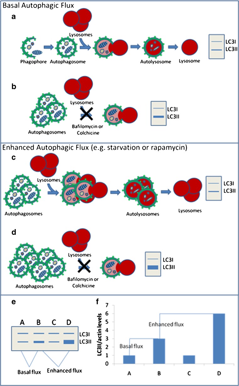
Fig. 2.
How to measure basal and induced autophagic flux. a An intact autophagic system produces and degrades LC3II/autophagosomes. b Blocking LC3II/ autophagosomes with compounds like BafA and colchicine reflect the production of LC3II in the cell or “flux.” c Interventions that enhance flux increase LC3II/ autophagosome production and degradation; therefore, on an immunoblot, LC3II levels may not change. d Blocking LC3II degradation in the setting of enhanced flux reveals the true increase in LC3II production. e Example of immunoblot and densitometric graph of autophagic flux assays. Condition A compared with B reflects basal flux, whereas comparing B to D reflects the amount of stimulated or enhanced autophagic flux

