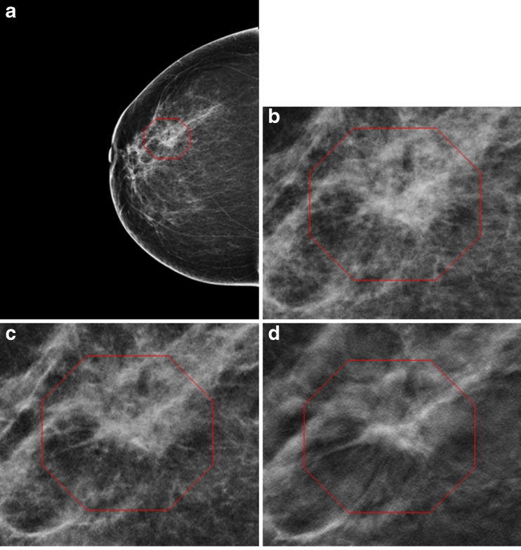
Fig. 3.
A 68-year-old woman. a Right breast cranio-caudal view (2D) shows a non-specific density. Enlargement of the 2D (b) and synthesised 2D (c) shows a suspicious but non-conclusive irregular density. On tomosynthesis (3D) cranio-caudal view, however, a spiculated mass consistent with invasive cancer is clearly seen (d). The cancer was missed by both readers in the 2D arm. Histology revealed an 8-mm invasive lobular carcinoma grade 2

