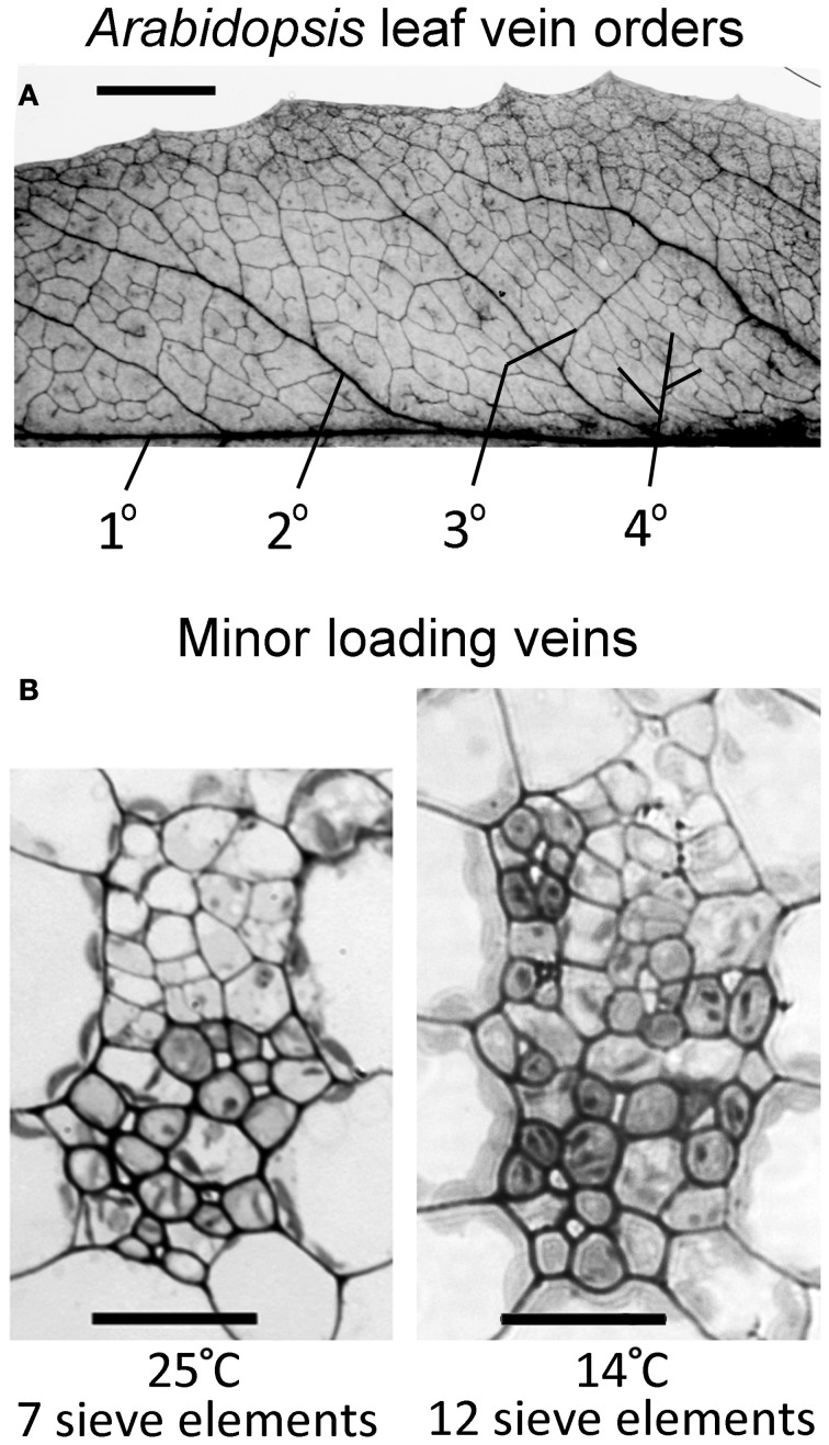
Figure 2.
(A) Vein orders in a cleared A. thaliana leaf (Swedish ecotype) grown at 25°C, with first order (1°) being the midrib and second-order veins (2°) comprised of main vascular branches off the midrib (typically five per leaf side, with five shown here either as portions or in their entirety), as defined by Hickey (1973). Third-order veins (3°) branch off the second order veins, and fourth-order veins (4°) are the smallest and can branch off all vein orders. (B) Cross-sectional light microscopic images are representative of fourth-order veins from mature leaves grown in warm (25°C) or cool (14°C) conditions under moderate light (400 μmol photons m−2 s−1). The vein shown from the plant grown at 25°C (B, left panel) contains seven sieve elements in the lower (abaxial) phloem region of the vein and features phloem parenchyma (angular and light colored) and companion cells (rounded and dark colored). The vein shown from the plant grown at 14°C (B, right panel) contains 12 sieve elements and features associated phloem cells encroaching into the upper (adaxial) region occupied by xylem cells. Scale bars shown are 5 mm for vein pattern and order (A) and 20 μm for fourth-order veins (B, same scale for both panels). The Italian and Col-0 ecotypes exhibited the same venation pattern and similar vein cross-sectional ultrastructure as shown here for the Swedish ecotype.

