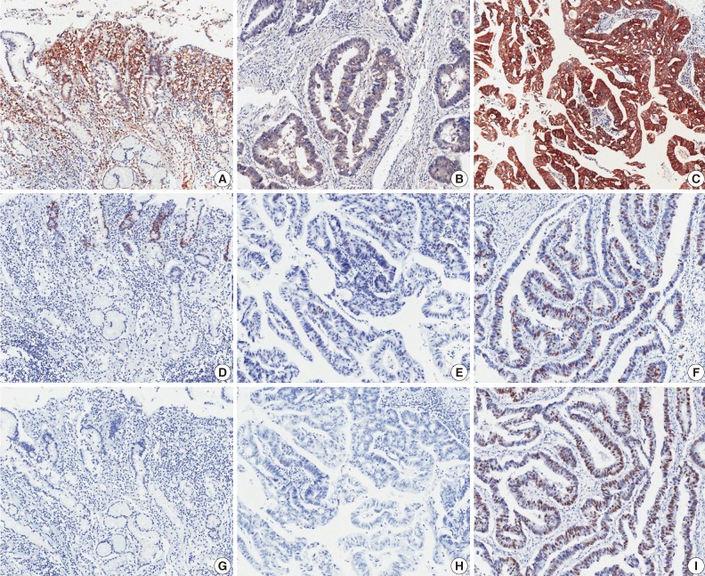
Fig. 1.
Microscopic images of immunohistochemical staining. Heat shock protein 70 (A-C), Ki-67 (D-F), and p53 (G-I). Left panels are from non-neoplastic gastric mucosa (A, D, G), middle panels are negative expression samples from gastric adenocarcinoma (B, E, H), and right panels are positive expression samples from gastric adenocarcinoma (C, F, I).

