Figure 6.
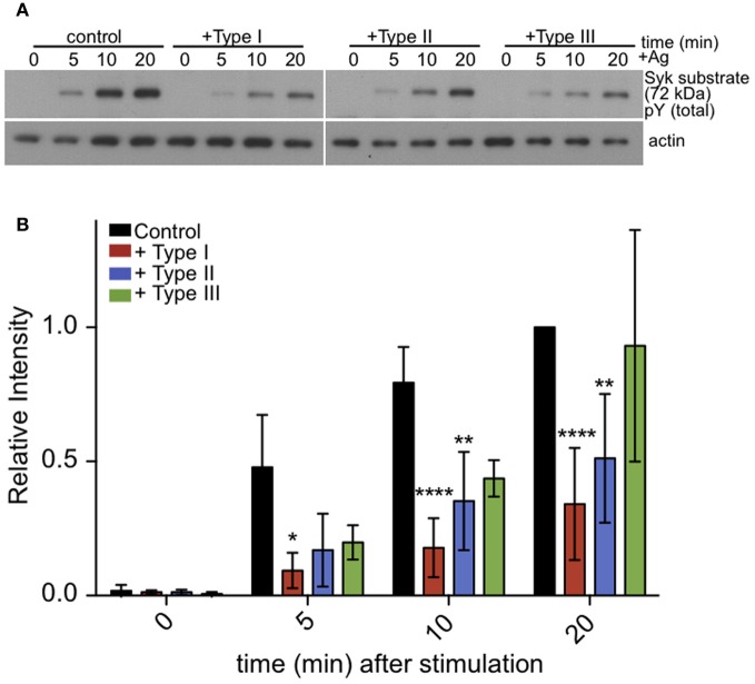
Tyrosine phosphorylation of pp72 Syk substrate is reduced in T. gondii infected cells. IgE-sensitized RBL cells were infected for 1 h with Type I, II or III tachyzoites as indicated. Antigen-stimulated cells were lysed at 0, 5, or 10 min after addition of multivalent antigen (10 ng/ml DNP-BSA). (A) Representative blot: Top panel shows phosphorylation of pp72 Syk substrate. Bottom panel shows loading control (α-tubulin) (B) Quantification of Syk substrate band intensity. Error bars represent SD of 4 independent experiments (*P < 0.05, **P < 0.01, and ****P < 0.0001 relative to uninfected cells).

