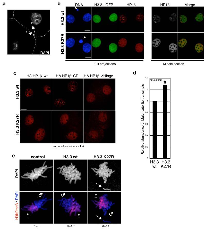
Figure 6. Expression of H3.3 – GFP K27R in the zygote leads to altered pericentromeric transcription, defects in chromosome segregation and mislocalisation of HP1β.
a. Embryos expressing H3.3 K27R display chromosome seggregation defects (24%; n =57) compared to embryos expressing H3.3 wt or H3.1 K27R or to non-injected embryos (5%, n=30; 8%, n=26 and 5%, n=21, respectively). Shown is a single section of a 2-cell stage embryo expressing H3.3 - GFP K27R stained with DAPI. White dashed line marks the cell membrane. Arrows point to DNA fragments that were not incorporated into the nuclei.
b. HP1β is mislocalised in 2-cell stage embryos expressing H3.3-GFP K27R. Zygotes injected with H3.3-GFP wt (n=19) or K27R (n=18) mRNAs were stained with an HP1β antibody. Shown are full projections (left) or middle sections (right) of images taken along the Z-axis every 0.6 μm. Embryos were processed in parallel and with identical confocal acquisition parameters. Non-injected embryos are shown in Supplementary Information, Fig. S5. Polar bodies are indicated by an arrow. Scale bar is 14 μm.
c. Deletion of the Hinge domain in HP1β results in mislocalisation of HP1β similarly to that observed upon H3.3K27R mutation. The indicated HP1β mRNA was co-injected with either H3.3 – GFP wt or K27R mRNA as in figure 1. Embryos were analysed with an anti-HA antibody. Shown are single confocal sections of representative 2-cell stage embryos. Scale bar is 14 μm.
d. Increased accumulation of major satellite transcripts upon H3.3K27R expression. Zygotes were injected with mRNA for H3.3 – GFP wt or K27R and cultured until early 2-cell stage. Total RNA was retrotranscribed and analysed by PCR with specific primers for major satellites. Data was normalised with H2A mRNA levels and is presented as average ± SD of 3 independent biological replicates. We observed no difference in major satellite transcription between H3.3 wt and non-injected embryos (not shown).
e. Paternal chromatin displays segregation defects during the 1st mitosis in embryos expressing H3.3 K27R. Non-injected (control), H3.3 wt or H3.3 K27R-expressing embryos were cultured till they reached mitosis, fixed and stained with a H3K9me3 antibody (which at this stage marks exclusively maternal chromatin). Paternal chromosomes (e.g. lacking H3K9me3) show severe division defects (arrow).

