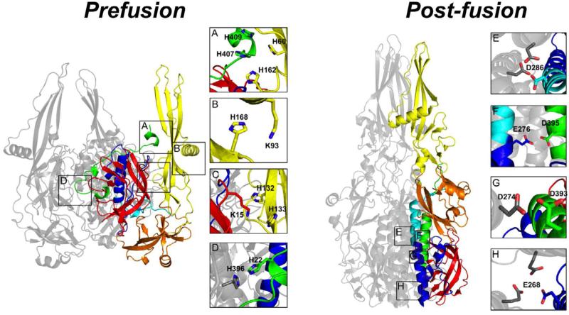
Figure 7. HisCat and AniAni interactions in VSV-G.
Prefusion) DI is red, DII is blue/green, DIII is orange, and DIV is yellow. DII can be divided into three sections that are non-continuous in the primary sequence, the NHR;blue, the MHR;Cyan, and the CHR;Green. In the compacted prefusion state a number of HisCat interactions are found, often between domains, and they are depicted in panels A-D (2J6J Biological assembly). Post-fusion) VSV-G elongates in the post-fusion state losing many of the HisCat interactions found in the prefusion state. In the post-fusion state many AniAni interactions form, occurring in DII and they are depicted in panels E-H (2CMZ).

