
Figure 5 .
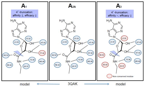
A comparison of the amino acids residues lining the subpockets of the ARs that are important for recognition of the ribose moiety. 3QAK is an X-ray structure of the 6 -A 2A AR complex, 20 which in this figure is adapted to 5 . The interactions of 3QAK were generalized by homology to A 1 AR and A 3 AR models (blue residues are conserved), and the general pharmacological effects of truncation are indicated. Many of these residues were detected as important in ligand recognition using site directed mutagenesis prior to the elucidation of the X-ray crystallographic structure.