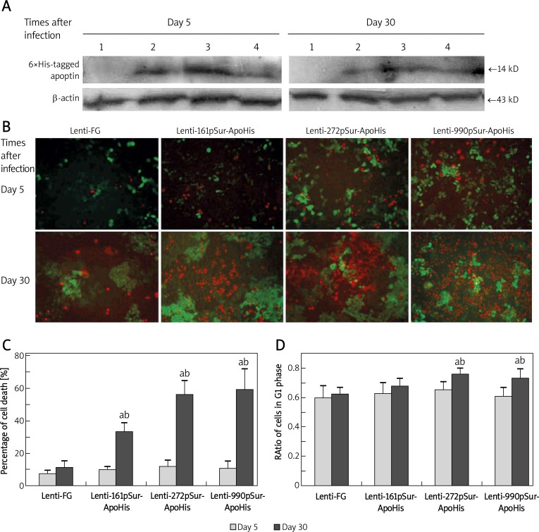
Figure 2.
The apoptin expression driven by different fragmental pSur in SW480 and its suppressive effect on the cancer cells. A – The expression of apoptin-6×His in SW480 cells detected by Western blot at 5 day and 30 day after the lentivirus infection: 1 – Lenti-FG; 2 – Lenti-990pSur-ApoHis; 3 – Lenti-272pSur-ApoHis; 4 – Lenti-161pSur- ApoHis. B – The apoptosis and necrosis revealed by dye binding under fluorescence microscope (bar = 100 µm) and C – the quantifying analysis of the dead cells. D – G1 phase ratio was examined by flow cytometry: a p < 0.05 vs. day 5 group;b p < 0.05 vs. control cells. All measurements were performed in triplicate

