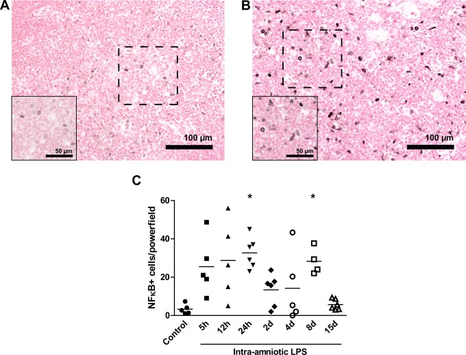
Figure 7.
Increased number of NF)-κB positive cells increased after LPS exposure.Representative images of NF-κB expression in the thymus are shown for controls (A) and 8-day LPS-exposed animals (B).The number of NF-κB positive cells in the medulla of the thymus increased 24 hours and 8 days after the exposure to intraamniotic lipopolysaccharide (LPS; C). *P < .05 versus controls.

