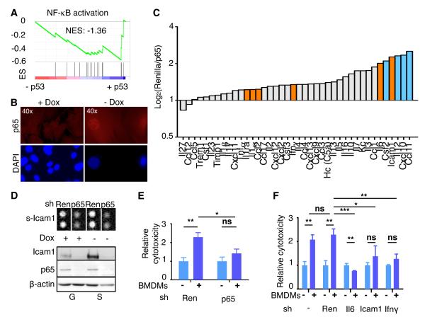
Fig. 6. NF-κB mediates p53-dependent SASP.
(A) GSEA plots evaluating p53-dependent changes in NF-κB signaling pathway.
(B) Immunostaining of proliferating (+Dox) and senescent HSCs (-Dox) with anti-p65 antibody (top panels) and counterstained with DAPI (bottom panels) to show nuclear NF-κB accumulation and SAHF formation, respectively, in senescent HSCs.
(C) Murine cytokine array for conditioned media from senescent HSCs transduced with Renilla or p65 shRNA. Bars represent the average of two independent experiments (log2). Factors upregulated in senescent HSCs (Fig. 4E) are indicated in orange.
(D) Top panel, cytokine array blot extract showing secreted Icam1 in cells transduced with Renilla or p65 shRNA, in proliferating or senescent HSCs. Bottom, p65 and Icam1 protein levels in the same cells. β-Actin serves as loading control.
(E and F) Cytotoxicity of senescent HSCs (blue) infected with shRNAs (Renilla and p65 in E; Renilla, Il6, Ifnγ and Icam1 in F) and incubated with (+) or without (-) BMDMs. The cytotoxicity (dark blue) is relative to the basal cytotoxicity (light blue, in the absence of incubation with BMDMs, which has been normalized to 1). Values are mean + SD from triplicates.
ES, Enrichment score. NES, Normalized enrichment score. Ren, renilla. BMDM, bone marrow-derived macrophages.

