
Figure 1. Representative CT scans of intracerebral haemorrhage.
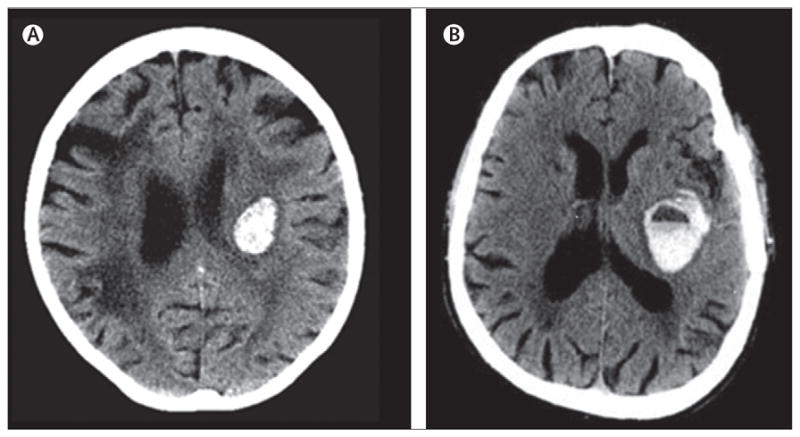
(A) This patient presented without medical history of anticoagulants. (B) This patient had raised international normalised ratio values and a positive history of vitamin K antagonist treatment at presentation. A fluid blood level is seen as a result of non-coagulated blood within the haematoma. (B) Reproduced from reference 24, by permission of the American Heart Association.