Figure 2.

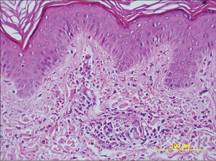
Skin biopsy specimen shows perivascular neutrophilic and lymphocytic infiltrate with leukocytoclasis and erythrocyte extravasation (H and E, ×400)

Skin biopsy specimen shows perivascular neutrophilic and lymphocytic infiltrate with leukocytoclasis and erythrocyte extravasation (H and E, ×400)