
Fig. 1.
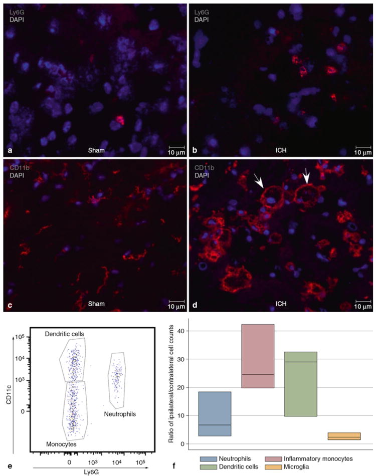
Immunohistochemistry of perihematomal brain post-ICH day 3 in an untreated mouse. (a) Ly6G staining (red) identifies rare neutrophils after sham surgery. (b) Increased numbers of infiltrating neutrophils are seen at the periphery of the hematoma. (c) After sham surgery, most CD11b+ (myeloid-derivative) cells are small and resemble resting microglia. (d) After ICH, CD11b+ cells have differing morphologies both within and at the periphery of the hematoma. Larger, vacuolated cells are indicated by the arrows. (e) Flow cytometry plot of CD45hiCD3-CD19-NK1.1-CD11b+ cells plotted Ly6G against CD11c to show three separate populations of dendritic cells (CD11c+Ly6G-), inflammatory monocytes (CD11c-Ly6G-), and neutrophils (Ly6G+). (f) Ratios of ipsilateral/contralateral cell counts in brain, n=3