
Figure 4. Tumor Trafficking of PrCSCs and hBM-MSCs to Human Cancer Xenografts in Mice.
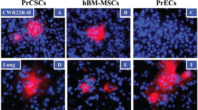
PrCSCs (A) and hBM-MSCs (B), but not PrECs (C), traffic to prostate cancer xenografts in vivo following systemic infusion. Fluorescently-labeled (CM-DiI, red) PrCSCs, hBM-MSCs, and PrECs (1 × 10⁶) were infused intravenously (IV) into immunocompromised mice bearing subcutaneous CWR22RH xenografts (3/group). Four days post-infusion, lungs and tumors were harvested and analyzed by fluorescence microscopy for the presence of CM-DiI-labeled cells. In contrast to the xenografts, all three cell types were found entrapped in the lungs following infusion (D-F). Nuclei counterstained with DAPI (blue). At least three images analyzed per tissue per animal, representative images shown.