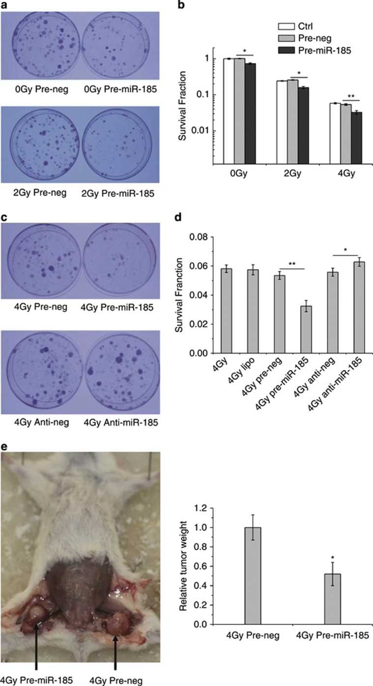
Figure 2.
Impact of miR-185 levels on cell survival. (a) Photographs of colonies formed by 786-O cells exposed to 0 and 2 Gy of X-rays. (b) Survival fractions of cells without transfection (Ctrl) and cells transfected with pre-miR-185 or pre-neg (30 nM final) in response to 0, 2 and 4 Gy of X-rays measured by colony formation assay. (c) Photographs of the colony formation with various treatments. (d) Survival fractions of 786-O cells with various treatments. (e) Tumor formation by the RCC cells in NOD/SCID mice subsequently exposed to 4 Gy of X-rays. Tumors formed by 786-O cells transfected with pre-miR-185 or pre-neg were separated from the hind legs of the mice (n=9) and weighed on the 55th day after subcutaneous injection. Each experiment was conducted at least three times independently. *P<0.05 compared with pre-neg; **P<0.01 compared with pre-neg

