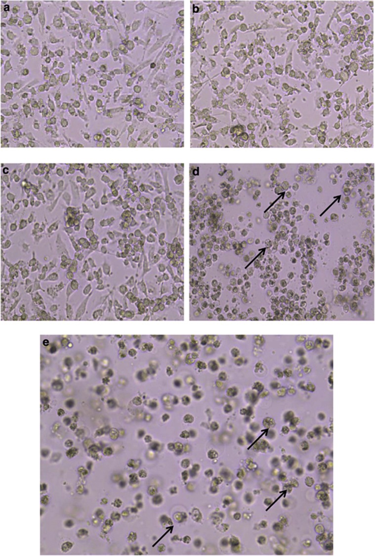
Figure 1.
HY-PDT apoptotic effects on cellular morphology of HepG2 human hepatocellular carcinoma cells. Cells were treated with increasing concentrations of HY (0.1, 0.2, 0.5 and 1 μg/ml HY) for 8 h followed by irradiation for 5 min. The cells were further incubated for 18 h and cellular morphology was examined for each treatment. (a) Cellular morphology of untreated HepG2 cells. The cells were aggregated and clustered as monolayer forms. (b and c) Early morphological changes during apoptosis in 0.1 and 0.2 μg/ml HY-PDT-treated cells, respectively. Cells were elongated and reduction of cell growth was observed. (d and e) Late apoptotic morphological changes in cells induced by 0.5 and 1 μg/ml HY-PDT compared with mock untreated cells. The cell confluency appeared to reduce from ∼90% in untreated to ∼20% in 1 μg/ml HY-treated cells. Apoptotic cells were detected as cell shrinkage and membrane blebbing. Similar cellular morphology was observed in three independent experiments (magnification × 100). The half-headed arrows represent apoptotic cells

