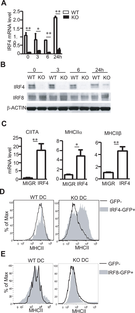
Figure 5. Contribution of decreased IRF4 expression to defective CIITA/MHCII expression in TSC1 KO BMDCs.
(A,B) IRF4 mRNA (A) and protein (B) levels in WT and TSC1KO BMDCs before and after LPS (10 ng/ml) stimulation for the indicated times. (C) upregulation of CIITA, MHCIIα, and MHCIIβ mRNA in TSC1KO BMDCs by IRF4 overexpression. TSC1KO BMDCs were similarly transduced with control MIGR vector or MIGR-IRF4 retrovirus as in figure 4C. CIITA, MHCIIα, and MHCIIβ mRNA levels in sorted TSC1KO GFP+ CD11c+ BMDCs were determined by real-time RT-qPCR. (D) Overlaid histograms show MHCII expression on GFP and GFP+ CD11c+ WT and TSC1KO DCs following transduction with IRF4 expressing retrovirus. (E) Overexpressed IRF8 couldn’t rescue MHCII expression in TSC1KO BMDCs. WT and TSC1KO BMDCs were similarly transduced with retrovirus expression IRF8 and analyzed for MHCII expression as in (C). **, p<0.01 determined by unpaired Student t-test.

