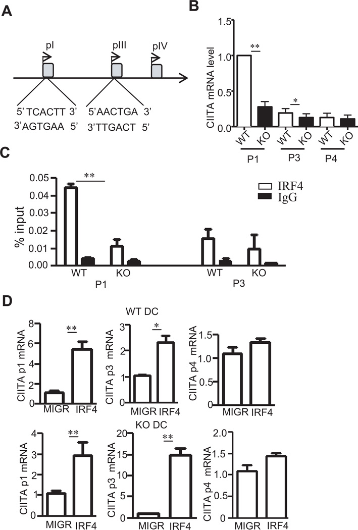
Figure 6. IRF4 binds to CIITA promoter1 and III.
(A) Schematic drawing of CIITA promoters. Potential IRF4 binding sites are shown. (B) CIITA pI, pIII, and pIV specific mRNA levels in WT and TSC1KO BMDCs determined by real-time qPCR. (C) Direct binding of IRF4 to pI and pIII of CIITA determined by ChIP and real time qPCR. WT and TSC1KO BMDCs were subjected to ChIP with anti-IRF4 or a control IgG, followed by real-time qPCR. The results are presented as immunoprecipitated pI and pIII relative to the total input DNA. Data were generated from three independent experiments. (D) IRF4 promotes pI and pIII but not pIV activity of CIITA in both WT and TSC1KO BMDCs. pI, pIII, and pIV specific CIITA mRNA in WT and TSC1KO BMDCs transduced with either GFP or GFP + IRF4 as described in figure 5C were quantified by real-time RT-qPCR. *, p<0.05; **, p<0.01 determined by unpaired Student t-test.

