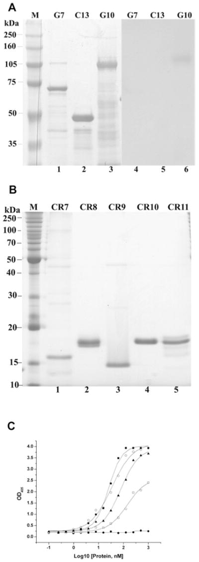
Figure 4. In toxin overlay assays and ELISA, the Cry11Aa toxin binds cadherin fragments and cadherin repeats.

(A) Partial cDNA fragments, G7, C13 and G10 (90 pmol each) were separated by SDS/PAGE (8 % gel) and stained using Coomassie Blue (lanes 1–3). These fragments were electotransferred to a PVDF membrane, which was incubated with 20 nM Cry11Aa toxin. Unbound toxin was removed by washing, the membrane was incubated with anti-Cry11Aa antiserum (1:2000) and then visualized by luminol (lanes 5–7). (B) The purified cadherin repeats CR7–11 (30 pmol each) were resolved by SDS/PAGE (15 % gel) and stained using Coomassie Blue (lanes 1–5). (C) The cadherin repeats, CR8 (○), CR9 (■), CR10 (□) and CR11 (▲) show dose-dependent binding to immobilized Cry11Aa (0.4 μg), but CR7 (●) does not bind Cry11Aa. OD405, A 405.
