
Figure 7.
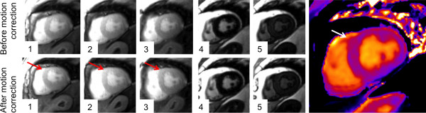
Failed motion correction during T1 mapping. The original images (upper row) show the regular shape of the LV myocardium (the first five out of eleven images of the complete T1 acquisition are depicted). The motion correction algorithm led to an outbound shift of the anterior and anteroseptal myocardial segment (red arrow in the bottom row). The corresponding map (right image) indicates an inhomogeneous T1 distribution in this area (white arrow).