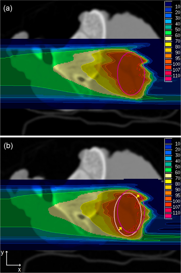
Figure 7.
Carbon ion plans of sample 2 with shifted PTV. The old PTV (light magenta) was shifted 2.0 mm in x- and 2.0 mm in y-direction to a new position (dark magenta): (a) adapted plan optimized on p CT after PTV shift and recalculated on r CT, (b) optimized on r CT before PTV shift and recalculated on r CT after PTV shift without plan adaptation. The unit of color scale is percent of the prescribed physical dose.

