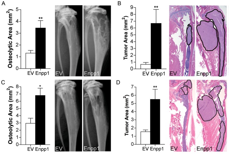
Figure 4. Effect of Enpp1 on the development of bone metastasis.
MDA-MB-231 cells stably expressing Enpp1 or empty vector (EV) were injected into the (A, B) tibia (n = 9/group) and (C, D) left cardiac ventricle (n = 5/group) of athymic nude mice and digital radiographic imaging was performed at weekly intervals. (A, C) Osteolytic area and (B, D) tumor area were measured on radiographic images and histological sections, respectively (*p<0.05, **p<0.01). Representative images are shown at 4 weeks following tumor cell administration. Black outlined areas on histological images indicate areas of tumor.

