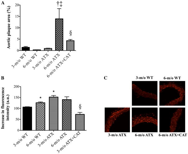
Fig. 3.
Effect of a 3-mo treatment with the antioxidant CAT (n = 6) on the atherosclerotic plaque area in the thoracic aorta (A) and the superoxide production quantified by dihydroethidium (DHE) staining (B) in aortic histological sections of 3- and 6-mo-old WT (n = 4 and 6, respectively) and ATX (n = 5 and 6, respectively) mice. C: representative images of DHE staining in the different groups of mice. Data are means ± SE of n mice. One-way ANOVA: P < 0.05 vs. 3-mo-old WT (*), vs. 3-mo-old ATX (†), vs. 6-mo-old WT (‡), and vs. 6-mo-old ATX (§).

