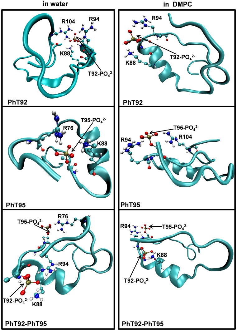
Figure 10. Structure of phosphorylated α2-peptides at the end of MD experiments in water and in DMPC.
The dimyristoylphosphatidylcholine (DMPC) membrane bilayer and water molecules are not shown in order to improve clarity. The images are a representative snapshot of singly-phosphorylated (PhT92, PhT95), and doubly-phosphorylated (PhT92–PhT95) α2-peptides (S72–S107) of myelin basic protein (MBP) captured during the last 10 ns of the 160 ns molecular dynamics simulation experiments. Shown are examples of the type of electrostatic interactions between phosphorylated Thr residues and different basic residues within the α2-peptide variants.

