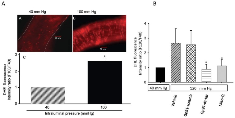
Figure 1. Elevation of intravascular pressure induced changes in superoxide production as determined by fluorescent microscopy analysis.
(I) An increase in intravascular pressure induced increased production of O2 – in cannulated rat cerebral arterial segments as revealed by a significant increase in the fluorescent intensity ratio of dihydroethidium (DHE) fluorescence (A vs B). The magnification in panel A or B is 200×. Panel C depicts bar graphs comparing the DHE fluorescence intensity ratio following pressurization of cannulated cerebral arterial segments at 40 and 100 mm Hg. The increase in intraluminal pressure from 40 to 100 mm Hg significantly increased the DHE fluorescence intensity ratio as summarized in panel C. *P<0.05, n = 3–5 independent trials. (II) in further studies, pressurization of the cannulated cerebral arterial segments from 40 mm Hg to 120 mm Hg markedly increased the DHE fluorescence intensity ratio, which was significantly attenuated by prior treatment with the peptide inhibitor of NADPH-oxidase gp91-ds-tat (5 µM) or with the mitochondrial antioxidant MitoQ (1 µM), whereas pretreatment with the control gp91-scarmbled-tat peptide had no effect. These findings indicate that NADPH oxidase and the mitochondria are the possible sources for the pressure induced ROS production (n = 4–5 independent studies for each group, *denote significant difference from control at P<0.05).

