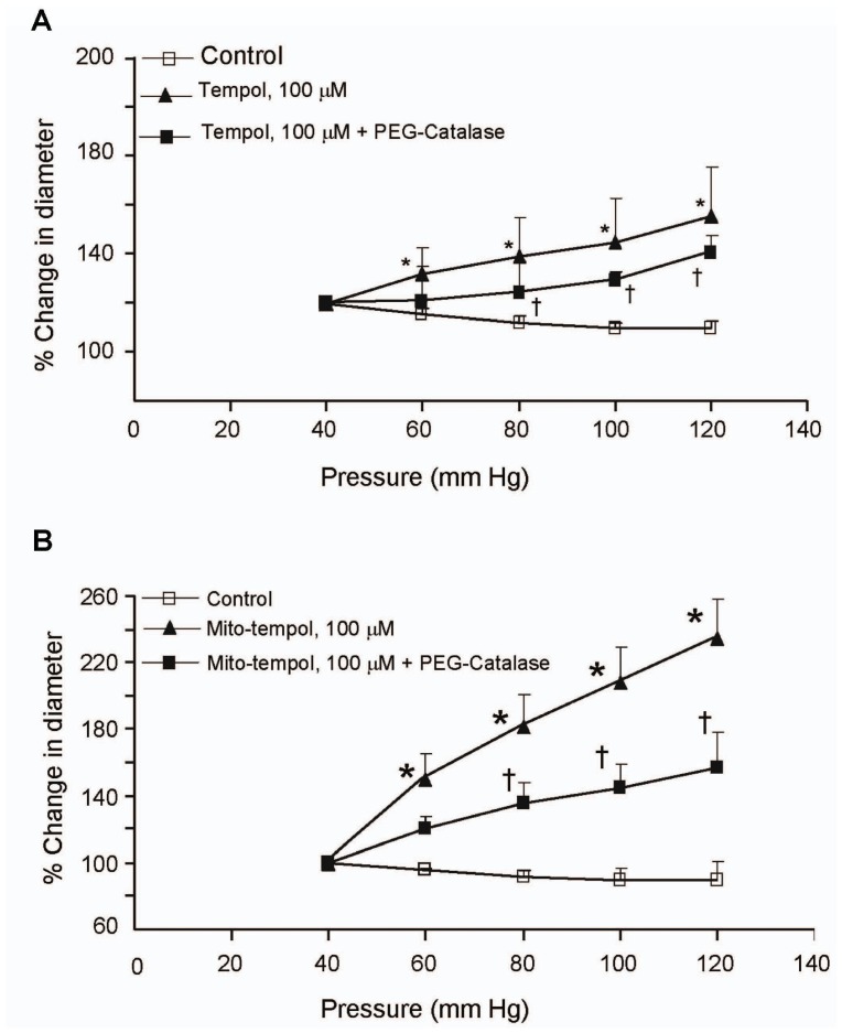
Figure 3. Dismutation of the ROS superoxide and H2O2 attenuated the increase in intraluminal pressure-induced myogenic constriction of cerebral arterial segments.
Increasing intraluminal pressure over the range of 60 mm Hg to 120 mm Hg in steps of 20 mm Hg induced pressure-dependent myogenic cerebral arterial constriction that was significantly attenuated and converted to vasodilation by pretreatment of the cannulated pressurized cerebral arterial segments with the superoxide dismutase mimic tempol and tempol plus the H2O2 dismutase PEG-catalase (A) or with mitochondrial targeted mito-tempol and mito-tempol plus PEG-catalase (B). Data are presented as mean value ± SEM, n = 6–8 cerebral arterial segments per group. *P<0.05, **P<0.001.

