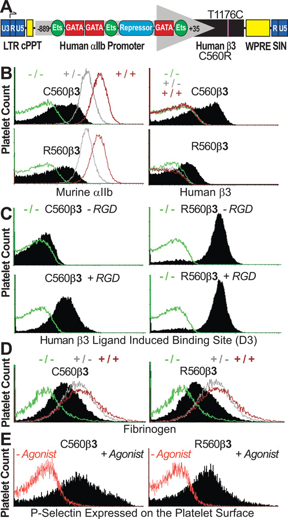
Figure 1. C560β3 & R560β3 tx Recipient Platelets Expressed αIIbβ3.
(A) β3-WPT Diagram. Viral 3'-long terminal repeat (LTR) enhancer/promoter was removed to self-inactivate vector (SIN) and αIIb gene promoter (nucleotides −889 to +35) directed megakaryocyte-specific transcription of cDNA with either nucleotide C1776T encoding a Cys (C) or Arg (R) at amino acid 560 of human β3. αIIb promoter binds GATA and Ets for high-level gene transcription in megakaryocytes while a repressor inhibits gene transcription in other lineages. The woodchuck hepatitis virus postregulatory element (WPRE) and central polypurine tract (cPPT) enhance transgene expression.
(B) αIIbβ3 was Detected on Platelets Tranduced with C560Rβ3. Left, immunocytometric analysis showed the mean fluorescence intensity (MFI) for αIIb(+) platelets in C560Rβ3 mice (black) appeared at moderate levels compared to β3(−/− green; +/− grey; +/+ brown) controls. Right, only C560Rβ3 tx mice displayed detectable levels of human β3 on platelets (black) compared to MFI levels of platelet controls. Results represent 15 experiments analyzing platelets from β3(−/−,+/−,+/+) controls and 23 C560β3 mice and 20 R560β3.
(C) αIIbR560β3 was Activated Continuously. Left, immunocytometric analysis revealed that C560β3 platelets (shaded peak) bound an Ab “D3” (recognizing an epitope exposed on high-affinity conformation of human β3 bound to ligand) only in the presence of a fibrinogen mimetic peptide containing Arg-Gly-Asp (+RGD). Right, in contrast, R560β3 platelets bound “D3” in the absence (−RGD) and presence (+RGD) of the peptide indicating that αIIbR560β3 was in an activated conformation. Negative control β3(−/−) platelets (green) failed to bind “D3”. Results represent four experiments using platelets from β3(−/−,+/−,+/+) controls and three C560β3 and R560β3 mice.
(D) Immunocytometric Analysis of Fixed/Permeabilized Platelets. Fibrinogen was absent from β3(−/−) platelets, although β3(+/−;+/+) controls displayed appreciable levels of platelet fibrinogen. C560Rβ3 platelets bound, endocytosed, and stored fibrinogen (shaded peak). Results represent ten experiments using β3(−/−,+/−,+/+) controls and three C560β3 and R560β3 mice.
(E) Immunocytometric Analysis Detected Activation of Platelets Treated with a mixture of ADP, epinephrine, and PAR4. The α-granule protein, P-selectin, was detected on the surface only after activation of C560β3 and R560β3 platelets (shaded peak) demonstrating that platelets expressing either form of β3 circulated normally in a quiescent manner. Results represent three experiments using platelets from β3(−/−,+/−,+/+) controls and three C560β3 and R560β3 mice.

