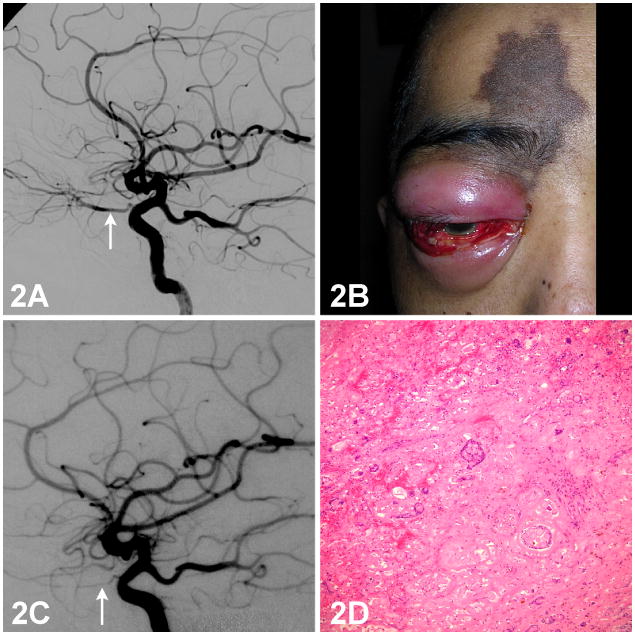
Figure 2. Internal carotid artery angiogram, external photograph of the eye, and photomicrograph of a histopathological section of the lacrimal gland from a patient with lacrimal gland adenoid cystic carcinoma.
2A: Diagnostic internal carotid artery angiogram showing tumor remnants supplied by the ophthalmic artery. (Arrow)
2B: Skin hyperpigmentation over the medial right brow and forehead. Anterior segment examination showing conjunctival injection, chemosis, hemorrhage, corneal edema, shallow anterior chamber, and hypotony.
2C: Repeat angiogram shows a hypoplastic right ophthalmic artery. (Arrow) Collateral flow from the middle meningeal artery and the distal maxillary artery of the external carotid artery (ECA) is present.
2D: Histopathologic examination of an exenterated specimen shows foci of tumor necrosis and blood vessel thrombosis. H/E 100X.

