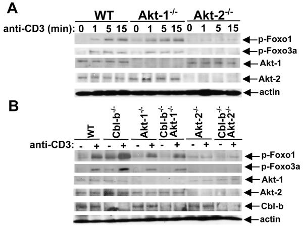
FIGURE 5.
Akt-2 but not Akt-1 phosphorylates Foxo1 and Foxo3a downstream of Cbl-b. Naïve CD4+CD25− T cells from WT, Akt-1−/−, and Akt-2−/− (A), or WT, Cbl-b−/−, Akt-1−/−, Cbl-b−/−Akt-1−/−, Akt-2−/−, and Cbl-b−/−Akt-2−/− mice (B) were stimulated with anti-CD3 for times indicated and lysed. The cell lysates were blotted with phospho-Abs against Foxo1 and Foxo3a, and reprobed with anti-actin. The presence or absence of Akt-1, Akt-2, and Cbl-b was determined by immunoblotting with anti-Akt-1, anti-Akt-2, and anti-Cbl-b, respectively. The data shown are one representative of three independent experiments.

