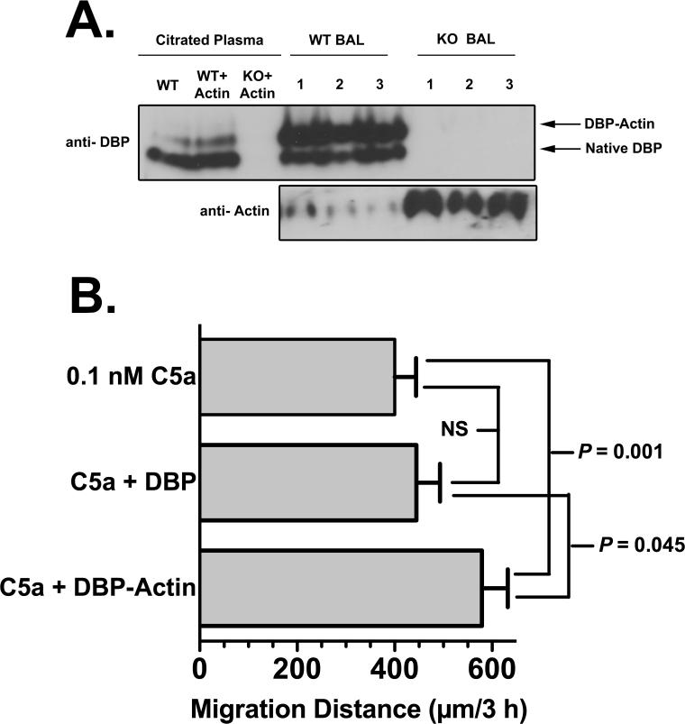
Figure 7.
DBP complexes with actin in BAL fluid. (A) Cell-free BAL fluid from DBP+/+ wild-type (WT) and DBP-/- knock-out (KO) mice obtained 4 hours after instillation of 1 μg purified mouse C5a was analyzed by native (non-denaturing) polyacrylamide (8%) gel electrophoresis. Complex formation in pooled citrated plasma from WT and KO mice spiked with 1 μM purified actin (this concentration will bind 15-20% of the total plasma pool of DBP) was included as a reference marker. Upper panel is an immunoblot with chicken anti-human DBP, lower panel is an immunoblot of the same BAL samples using pan anti-actin mAb (clone ACTN05). Note that when actin is bound to DBP it is not detected by the anti-actin mAb, only unbound actin is detected. Numbers indicate BAL samples from individual WT or KO mice. (B) Chemotaxis of normal human neutrophils to purified C5a (0.1 nM), C5a + 1 μM purified DBP, or C5a + 1 μM purified DBP-actin complex (n = 6). Chemotaxis was performed using the under agarose method for 3 h at 37°C. Statistical significance is indicated.

