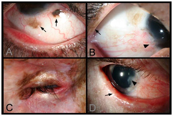
Figure 2. Lid and ocular surface manifestations in Xeroderma pigmentosum (XP) patients.
A) Conjunctival melanosis (arrows) in Case 5, 8-year old Asian Indian XP-C patient (XP417BE). Note the feeder vessels to lesions (arrows). B) Early pterygium (arrowhead) and lid pigmentation (arrow) in Case 2. C) Severe ectropion, entropion, and ocular inflammation in Case 3. D) Lid margin keratinisation (arrow), loss of lashes in Case 6, a 14 year old patient (XP243BE). The patient has a history of skin cancer but no history of ocular surface cancer. Lentigines are present on eyelids and patient has bilateral pterygium, and ectropion. Patient has decreased best corrected visual acuity, possibly due to amblyopia. Localized corneal clouding at leading edge of pterygium was suspicious for early malignancy, biopsy recommended.

