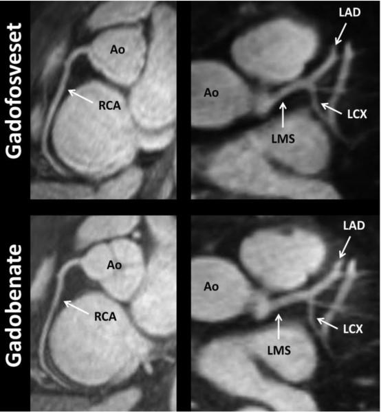
Fig. 1.
22-year-old male healthy volunteer. Good quality representation of same individual with both contrast agents. The arrows indicate the general locations of the ROIs that were measured at the proximal sections of the various arterial segments. Note – Ao, Aorta; LMS, left main stem; LAD, left anterior descending coronary artery; LCX, left circumflex coronary artery; and RCA, right coronary artery.

