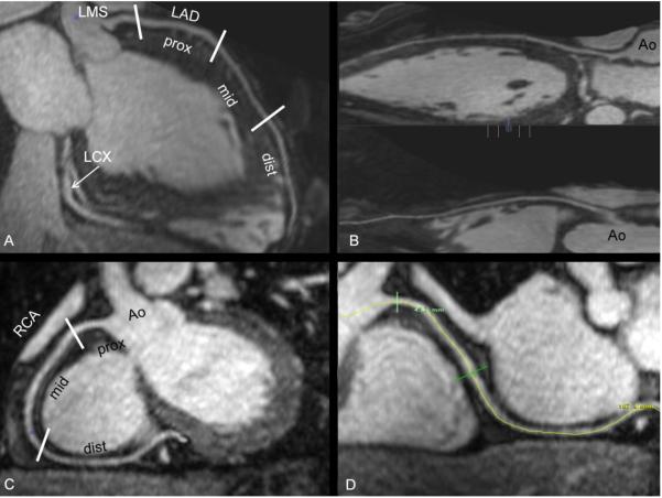
Fig. 2.
27 year-old woman healthy volunteer. Study done with Gadovosveset. (A and C) Segment analysis of LAD and RCA. (B and D) Reformatted images through curve maximum intensity projection (MIP) of LAD and RCA, respectively. Note – Ao, Aorta; LMS, left main stem; LAD, left anterior descending coronary artery; LCX, left circumflex coronary artery; and RCA, right coronary artery. Prox, mid and dist indicates proximal, middle and distal coronary segments, respectively.

