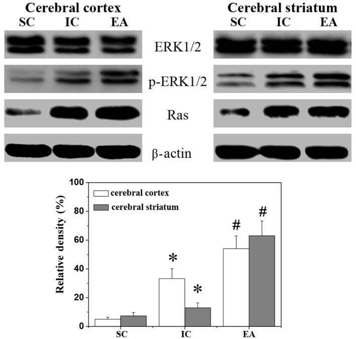
Figure 2.
Effect of EA on the ERK1/2 pathway in cerebral I/R-injured rats. The levels of ERK1/2 protein expression and ERK1/2 phosphorylation in the ischemic cerebral cortex and cerebral striatum were determined by western blotting. β-actin was used as the internal control. Relative density was expressed as the optical density of p-ERK1/2 relative to that of tERK1/2. Data are representative of five individual rats in each group. Data are averages with SE (error bars). *P<0.05, vs. SC group; #P<0.05, vs. IC group. I/R, ischemia/reperfusion SC, sham-operated control; IC, ischemic control; EA, electroacupuncture; ERK, extracellular signal-regulated kinase.

