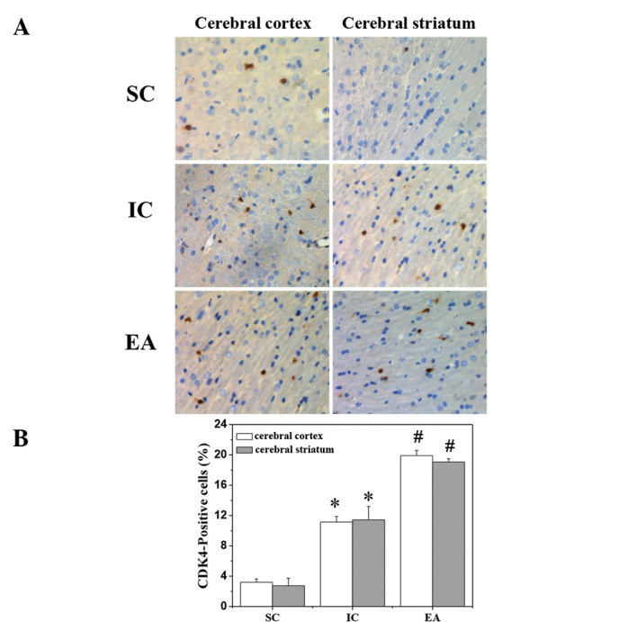
Figure 5.

Effect of EA on the CDK4-positive cell rate in cerebral I/R-injured rats. Cerebral tissues from each group (n=5) were processed for an immunohistochemical assay. The nuclei of all cells were visualized by hematoxylin staining and the CDK4-positive cells were stained sepia with DAB solution. CDK4-positive cells were counted at four randomly selected microscopic fields ×400 magnification. The CDK4-positive cell rate was expressed as the ratio of sepia-stained cells to the blue-stained total cells. Data are averages with SE (error bars). *P<0.05, vs. SC group; #P<0.05, vs. IC group. SC, sham-operated control; IC, ischemic control; EA, electroacupuncture; CDK4, cyclin-dependent kinase 4; I/R, ischemia/reperfusion; DAB, diaminobenzidine.
