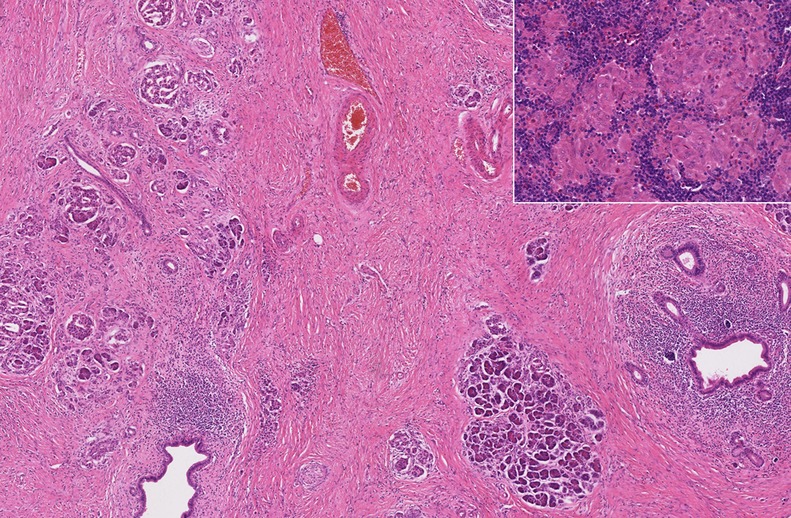
Figure 2.

Medium power histological view of pancreatic resection specimen sample showing severe fibrosis, atrophy of pancreatic parenchyma and duct-centred chronic inflammation, with retention of a normal lobular architecture (H&E). Inset: high-power histological view of small epithelioid non-caseating granulomata within peripancreatic lymph nodes, consistent with sarcoidosis (H&E).
