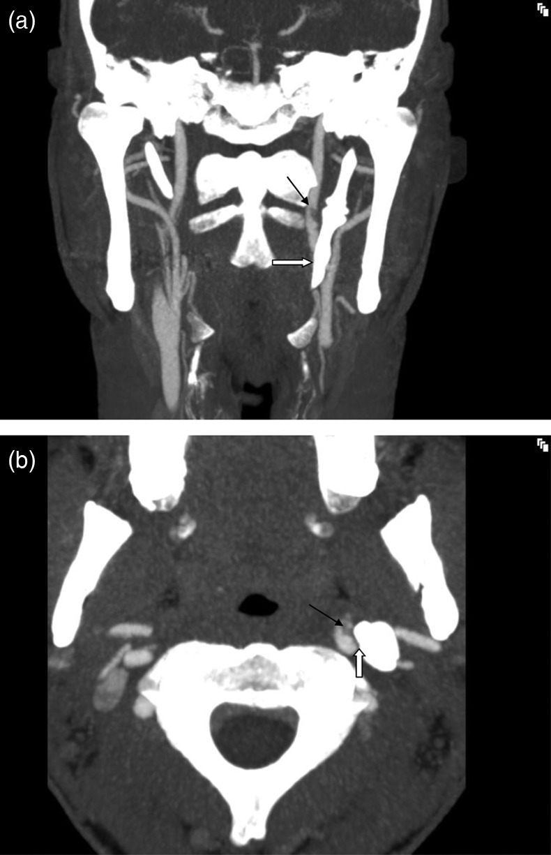
Figure 1.

A CT-angiography with coronal view of the neck shows an elongated styloid process (SP) (white arrow) in close proximity to the internal carotid artery (ICA) on the left side. Note the filling defect from the clot in the vessel (black arrow). (B) CT-angiography with axial view of the neck shows the close contact between the SP and the ICA on the left side (white arrow). There is a clot in the vessel due to dissection (black arrow).
